When you see a bluish vein popping up on the surface of your skin, it’s likely you’d recognise they are varicose veins. But is that all of them? Are there any you can’t see?
When you go to get your varicose veins or spider veins treated, it’s vital that you get a complete examination and diagnosis. It’s not enough for a health professional to only take your medical history or do a visual examination of the problem veins. Varicose veins usually do not spontaneously arise on the surface of the skin, there are usually internal veins that you can’t see which feed the surface vein. Many patients’ venous insufficiency starts high up in the thigh or groin, which compound backward pressure causing the veins you see on the skin surface.
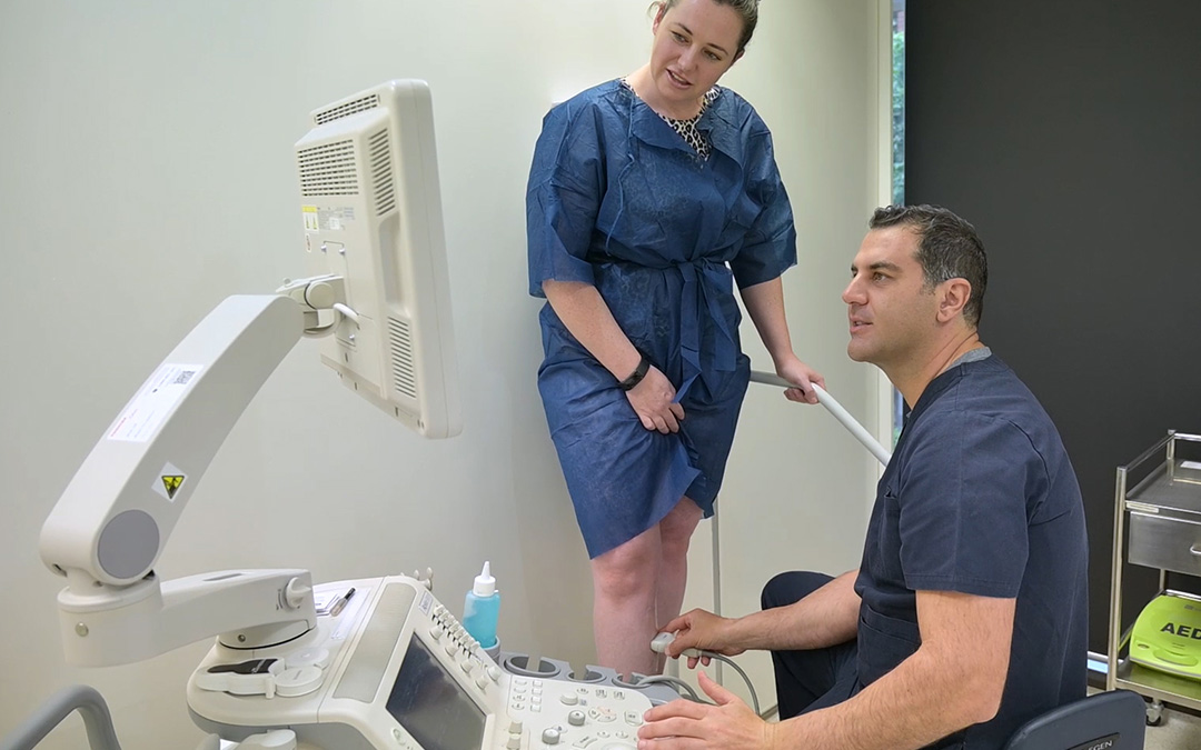
An ultrasound scan allows your phlebologist to not only identify the veins that may be causing you direct pain and symptoms – it also allows them to trace the origin of the problem veins. If your doctor does not address these feeding veins in your treatment, before long, you will develop more varicose veins in that region of your leg. In fact, this is the most common cause of varicose vein recurrence – overlooking treatment of the feeding veins.
Spider veins need ultrasounds too.
It’s common for people who have visible spider veins on the legs, but no larger varicose veins, to seek a cosmetic procedure (such as IPL laser) to treat what they assume is a superficial cosmetic problem. However, spider veins on the legs are often an indicator of underlying unseen vein problems below the surface of the leg.
Spider veins on the legs are usually the “overflow” of varicose veins that are so incompetent at returning blood to the heart that the excess pooled blood needs somewhere to go. This forms tributaries of smaller veins. An ultrasound diagnostic shows where the tiny veins originate, and importantly, the underlying veins can be treated instead of overlooked. Failing to treat the underlying incompetent veins may lead to spider vein matting later (a cluster of extremely find spider veins).
The importance of having a high-end ultrasound machine
Ultrasound technology has come a long way since its invention and introduction to the medical profession in 1949 by English physicist John Wild. An ultrasound machine uses sound waves emitted from a probe to provide images on a computer screen. The better the ultrasound unit and probe, the sharper and more defined the images produced. This makes it ideal for tracing fine tributaries of fine veins and varicose veins to their origins.
Here’s why state of the art ultrasound machines, which provide the best quality imaging, are important in leg vein treatment:
Diagnostic tests
Better images allow the treating phlebologist to identify all sources of reflux which would have previously been missed using older ultrasound units. If the abnormal veins are correctly identified this helps the phlebologist employ the right treatment, providing a more successful outcome for the patient.
Accurate ultrasound reports
Both the sonographer and phlebologist should have specialised training specifically in vascular sonography to ensure detailed and accurate ultrasound reports. With the help of the latest ultrasound machines, ultrasound reports can be highly precise and consistent.
Safety during treatment
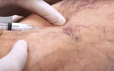
Safety is extremely important when treating varicose veins. High-quality ultrasound imaging makes it easier for the treating phlebologist to guide the sclerotherapy needle or catheter into the right vein, then confirm and target treatment. If treatments are incorrectly inserted into arteries instead of veins, this can be dangerous to the patient by sealing off active arteries carrying oxygen to the extremities, rather than sealing useless incompetent veins attempting to return deoxygenated blood to the heart.
Accreditation
It is now mandatory for all radiology and ultrasound departments to be nationally accredited. This also applies to phlebology practices that provide ultrasound diagnostic services. Vein Health Medical Clinic has maintained its Diagnostic Imaging Accreditation (DIAS) and will continue to do so.
Key Takeaways
- Ultrasound examination is an essential tool in the diagnosis and treatment of varicose veins and spider veins.
- Ultrasound scans are used to trace the origin of the varicose vein and spider veins in the leg, which may be high up in the thigh or groin, invisible to the patient.
- High-end ultrasound machines have excellent resolution and provide clear images to guide treatment, generate accurate imaging, and improve patient safety.