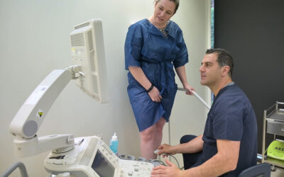
Patients often ask me “Why do I need an ultrasound examination to assess my varicose vein problem?” The answer to this is simple. Most Surface veins are connected to larger and deeper veins. These hidden veins, if abnormal, act as direct feeders, pushing blood in the wrong direction and into the bulging surface veins. The phrase I often refer to is “The Tip of the Iceberg”.
Hidden veins may include the well-known saphenous veins or abnormal vein connectors referred to as perforators.
The only way to examine these deeper veins is to use an ultrasound. Ultrasounds use sound waves that are transmitted into the leg and bounce off soft tissue structures to create a black and white image.
This technology dates back to World War I, when sonar was used to detect submarines deep in the ocean. Modern-day ultrasound units also use colour to determine the direction of blood flow within varicose veins. This helps delineate a normal vein from an abnormal vein.
Without an ultrasound examination, we can never really fully determine the extent and underlying severity of varicose veins.
 As time has passed, ultrasound machines have become more advanced, with the ability to deliver an incredible resolution, consequently improving the accuracy and efficiency of the examination process.
Patients are assured that at Vein Health Medical Clinic, the latest and most advanced Toshiba Ultrasound Units (Toshiba Aplio) are used, upgraded, and maintained. This ensures that the diagnostic procedures are world-class and conform to the standards required for a modern-day Phlebology Practice. In addition, we have specialised sonographers who are specifically trained to use these advanced machines and provide accurate reports.


