Spider Veins
and Reticular Veins
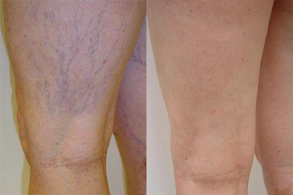
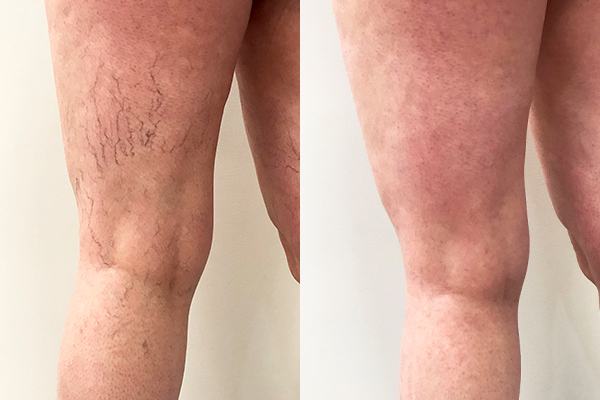
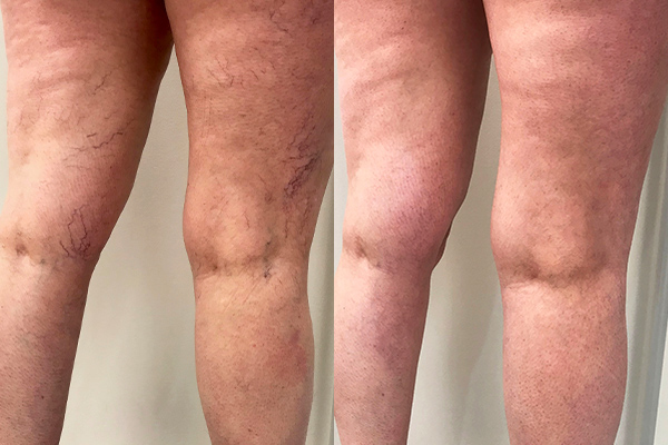
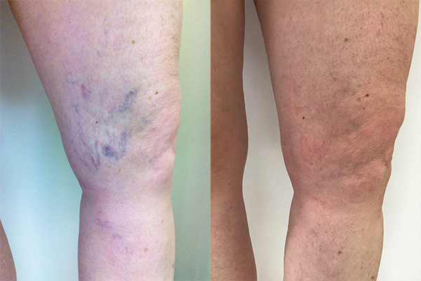
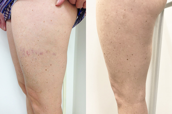
Spider veins are the tiny red or blue veins that may appear anywhere on the body, but more often on the legs, ankles, face, chest, or abdomen. They are extremely common. Spider veins may appear to be short, seemingly unconnected lines, each about a hair’s width, or they may resemble a spider web or tree with branches. In medical terms spider veins are called telangiectasias and usually occur in association with larger dilated blood vessels (often blue / green in colour) called reticular veins. Dilated blood vessels may cause aching especially with prolonged standing.
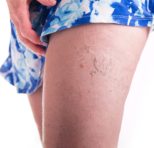
What causes spider veins?
Spider veins are often a flow-on effect of varicose veins in nearby blood vessels. When the damaged section of vein (varicose vein) fills with blood and swells to its capacity, the overflow of blood creates tiny spider veins close to the skin. This is why it is important to treat the underlying cause, and not just the spider veins that can be seen on the skin surface.
Are spider veins dangerous?
Generally speaking, the answer is no. However, if spider veins are a result of underlying venous disease, then the underlying condition causing the spider veins must be treated, as it could lead to further complications if left untreated.
Do you need these veins?
Although dilated blood vessels do carry blood they are not efficient and are often not necessary to the circulatory system. The body will have already established an alternative route for the blood to travel back more efficiently to the heart. Thus, they can be treated without a negative effect on circulation. Treatment will actually improve circulation.
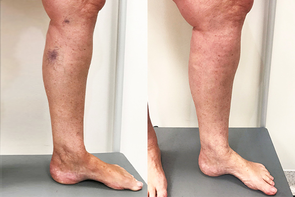
There is an approximately 80% chance for a greatly improved appearance with sclerotherapy, depending on the severity of the problem. Treatment will also usually relieve any symptoms caused by the veins.
Spider veins appearing in the lower leg, ankle or foot can be a sign of serious varicose vein disease. Spider veins in those areas are almost always fed by a larger internal vein that is malfunctioning and could lead to further complications if left untreated.
How do phlebologists classify the severity of venous disease?
In the past, it has been tricky for phlebologists and health professionals to define vein disorders because of the range and severity of symptoms. By creating the CEAP venous disease classification system, discussion of conditions and treatments is standardised across the world. Read more about the CEAP classification system.
How We Treat Spider Veins and Reticular Veins
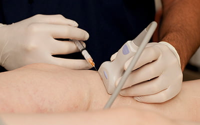
Ultrasound Guided Sclerotherapy
Ultrasound Guided Sclerotherapy is a highly specialised procedure involving injecting a sclerosant solution into the abnormal veins using ultrasound guidance, causing the vein wall to collapse. The veins dissolve and disappear as the body gradually absorbs them.
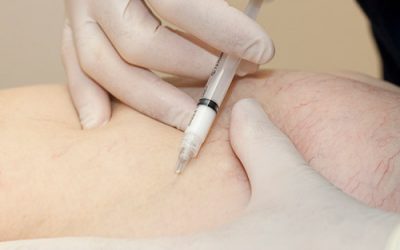
Sclerotherapy
The gold standard treatment of spider veins is sclerotherapy, also known as microsclerotherapy or direct vision sclerotherapy. It involves the injection of an irritating solution, through a tiny needle into the spider veins which causes them to close over and harden over.